

 FP604 : Novel Panoramic OCT Angiography: The Non-Invasive Imaging Tool FOR CENTRAL SEROUS CHORIORETINOPATHY
Dr.Rakesh Juneja ,Dr.Manish Nagpal,Dr.Navneet Mehrotra,Dr. Ahmed Eltayib
Introduction:
Central serous chorioretinopathy (CSC) is a disorder characterized by episodes of macular serous retinal detachment (SRD), first described and characterized as a clinical entity with fluorescein angiography (FA) [1–3]. Several risk factors have been identified, such as glucocorticoid levels, type A personality, pregnancy, uncontrolled systemic hypertension, use of antibiotics, bone marrow or organ transplantation, infection of the respiratory tract, and infection by Helicobacter
pylori [4–11]. The disease may present with different patterns: acute CSC, persisting, recurrent, and chronic CSC [12]. It usually occurs in young males but may be observed in older subjects of both genders. CSC is characterized by
retinal pigment epithelium (RPE) changes that are easily observed on fundus autofluorescence (FAF) pictures, serous retinal pigment epithelium detachment (PED) and episodes of SRD [12]. FA usually shows areas of fluorescein leakage,
often focal, with a smokestack or inkblot pattern in acute CSC [1–3], and more widespread in chronic stages, also called diffuse retinal pigment epitheliopathy [1, 3]. Indocyanine green angiography (ICGA) may show a hyperpermeability
of the choroidal vessels, and sometimes diffuse dye leakage [13]. Optical coherence tomography (OCT) and especially enhanced-depth imaging (EDI) or swept source (SS) OCT shows a thick choroid and may allow better characterizing the areas of PED and SRD [14, 15]. FA however, has certain limitations as the dye is masked by retinal pigment epithelium (RPE) and gets leaked from choriocapillaris. ICG is also considered but being an invasive procedure it carries certain risks like brochospam, laryngospasm, anaphylaxis, cardiac arrest and is contraindicated in iodine allergy, hepatic disease, chronic renal failure patients on hemodialysis.
OCT angiography (OCTA) is a non-invasive tool that works on the principle of ‘decorrelation’ and results in the formation of en face images of retinal capillary network and choroidal vasculature without requiring dye. Angio Scan OCT Angiography software on the RS-3000 Advance OCT (Nidek) provides ‘panoramic images’ with larger fields of view (Figure 1). Panoramic OCTA (P-OCTA) images allow 3-D analysis, are repeatable on follow-up examinations and to a certain extent are comparable to FA and ICGA in terms of resolution. This study aims to describe a novel technique of finding leakage in Central serous Chorioretinopathy (CSR) by novel Panoramic imaging (P-OCTA) & to compare these findings with Fluorescein angiography (FA) & Indocyanine green angiography (ICGA). [16]
Material and Methods:
Study design and patient enrolment:
This is a single-center, prospective, cross-sectional, observational study approved by the Institutional Review Board. All cases provided informed consent to be enrolled in the study and data was collected from patients diagnosed as CSR at retina clinic from January 2018 to June 2018.
Eligibility criteria and data collection:
Data was collected under the following headings: demographic details, history of presenting illness, eye laterality, history of associated systemic co-morbidities, clinical examination details including visual acuity at presentation and follow- up with multimodal imaging features, treatment details, complications (if any) and treatment given for that complication. Inclusion criteria were: clinical findings consistent with acute, chronic, pre-treatment and post-treatment features of CSR and complete availability of documentation. If any one of the above-mentioned inclusion criteria was not met, patients were excluded from the study.
Treatment Regime:
Patients were either observed or treated with focal laser depending upon the location of leakage (on P-OCTA and FA).
Characteristic features of Lesion:
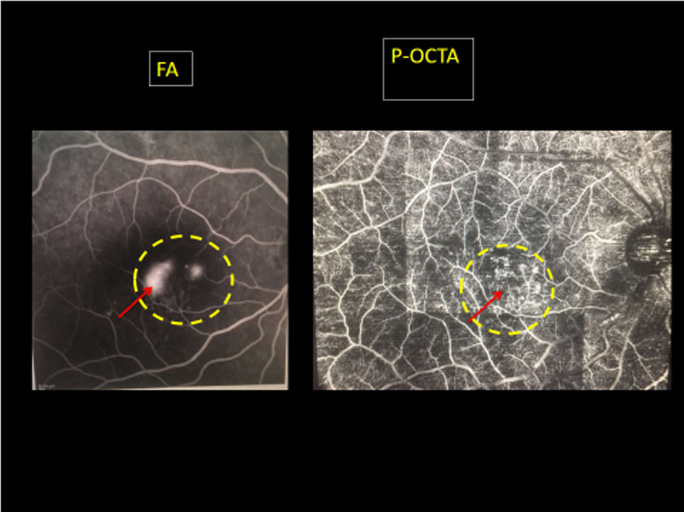
The acute lesions (pre-treatment) of CSR on P-OCTA appeared dilated, raised, well demarcated exactly at the same topographic location as of FA. Whereas the lasered ones and chronic lesions appeared flat, smooth also exactly at the same topographic location as of FA.
Study Parameters:
All cases underwent a complete eye examination at presentation and also at every follow-up visit. The examination protocol included best corrected visual acuity (BCVA) with the help of Snellen’s Chart, dilated fundoscopic examination, color fundus images (Topcon 50 Dx, Medical Systems Inc, Oakland, NJ, USA), fundus autofluorescence (FAF), fundus fluorescein angiography (FFA), indocyanine green angiography (ICGA) (Spectralis, Heidelberg Engineering Inc, Heidelberg, Germany), and spectral domain-optical coherence tomography (SD-OCT) (Spectralis, Heidelberg Engineering Inc, Heidelberg, Germany). Panoramic optical coherence tomography angiography (P-OCTA) (RS-3000 Advance OCT, Nidek) was performed in all eyes centered on the fovea covering 12×9 mm area (40° x 30° field of view). AngioScan RS-3000 Advance software was used to segment the outer retinal layers and choriocapillaris.
Panoramic Optical Coherence Tomography Angiography:
For all cases, at presentation and follow-up, P-OCTA images were obtained by Panorama feature of AngioScan OCT Angiography software on the RS-3000 Advance OCT (Nidek, Japan). This feature enables scanning a wide area—12 x 9 mm, 9 x 9 mm, 6 x 6 mm, and 4.5 x 4.5 mm thus providing large fields of view, 40° x 30°, 30° x 30°, 20° x 20°, and 15° x 15° respectively. In each of these sizes of fields of view, the software splits the entire selected scanning area into small fixed 3 x 3–mm cubes. The resolution obtained with a generated panoramic image is therefore the same as that of an individual 3 x 3–mm cube provided by other software, but a larger field is simultaneously scanned at the same time. The en face images obtained with panorama OCTA allowed to visualize individual retinal vascular plexuses and choriocapillaris. For all cases, P-OCTA scan covering an area of 12×9 mm equivalent to 40° x 30° field of view, was used at presentation and also on each follow-up.
Fluorescein Angiography and Indocyanine Green Angiography:
Corresponding to panoramic area (12×9 mm) imaged on OCTA with fovea as the very center, simultaneous and corresponding FA and ICGA images were obtained once adequate pupillary dilation (minimum of 7 mm) was achieved, at baseline (prior to initiating therapy) and at subsequent follow-up visits (on the discretion of the treating clinician). Sequential fundus images were obtained from early to late frames in all cases. Heidelberg Spectralis® (Heidelberg Engineering, Heidelberg, Germany) was used to acquire the angiography images.
Color Fundus Photography, Enhanced-depth Imaging Optical Coherence Tomography and Fundus Auto-fluorescence:
All cases at presentation and at each follow-up visit underwent a) Color fundus photography (Topcon 50 Dx, Medical Systems Inc, Oakland, NJ, USA) and a fundus mosaic was made in cases where lesions extended beyond arcades, b) SD-OCT (Spectralis®, Heidelberg Engineering, Germany) using the dense line-scan protocol with the EDI function activated. The minimum averaged number of frames per B-scan was 100, c) FAF imaging prior to FA/ICGA using Heidelberg Spectralis®.
Image Analysis:
For all cases, images were examined by two independent masked examiners. These examiners were ophthalmologists with subspecialty training in retina and uveitis (MN and NM). P-OCTA images were analyzed using the Angioscan software (RS-3000Advance). The software divided the retinal and choriodal vasculature into pre-defined slabs for segmentation (superficial and deep retinal vascular plexus, outer retina and choriocapillaris), however, manual segmentation was used to confirm the findings on P-OCTA. Images of only the choroidal vasculature generated on P-OCTA were compared, correlated and analysed with ICGA and FA images of corresponding region. This helped to assess whether P-OCTA scan can non-invasively help in detection of similar features as provided by invasive ICGA and FA.
Statistical Analysis:
Descriptive analysis was used to compare the appearance of the panoramic OCTA imaging with FA & ICGA
Results:
18 eyes of 18 patients were studied. The mean age at presentation was 35.8±11.6 years (range 13–62 years). All cases were males. All subjects included in the study were Asian Indians. Out of all cases included, 10 had acute, 8 had chronic presentation, 10 were pre-treatment and were 4 post-treatment cases. Few eyes included had lesions inside FAZ and few outside. All eyes had unilateral presentation. (Table 1)
Comparisons between Panoramic Optical Coherence Tomography Angiography, Indocyanine Green Angiography, and Fluorescein Angiography
15 eyes out of 18 patients with the clinical diagnosis of CSC showed an image pattern of areas of increased decorrelation signals (high signal intensity) in the OCT angiograms (P-OCTA) (Figures 2-6). Among the 15 eyes, 10 eyes additionally demonstrated dilated capillaries in the OCT angiograms. Among the 18 patients, leakage was observed in 14 eyes, whereas transmitted fluorescence and no obvious fluorescein leakage were observed in 2 eyes and 2 eyes, respectively. All eyes with CSC showed a choroidal hyperpermeability on ICGA. The areas that showed high signal intensity or dilated choriocapillaris in the OCT angiograms demonstrated a leakage in fluorescein angiography and hyperpermeability in indocyanine green angiography.
Comparisons between Panoramic Optical Coherence Tomography Angiography and routine 3 x 3 mm Optical Coherence Tomography Angiography
The various comparative features and differences between ‘panoramic’ OCTA to a routine 3 x 3 mm cube of OCTA have been described in Table 2
Discussion:
In our clinical study on patients with CSC, panoramic OCT angiography showed to be a valuable noninvasive tool to detect vascular abnormalities’ in choroid of eyes with clinical diagnosis of CSR. OCT-angiography revealed
a high intensity pattern in the choriocapillaris. The results of our study agree with previous investigations. Teussink et al [17] evaluated depth resolved
flow in patients with chronic CSC by OCT angiography and compared the findings with results obtained by fluorescein angiography and indocyanine green angiography. Examining 18 eyes with chronic CSC and 6 healthy controls, the authors reported that abnormal areas in the choroid were most often found in late-phase indocyanine green angiography and in OCT angiography of the choriocapillaris. Abnormalities in late-phase indocyanine green angiography and
fluorescein angiography co-located with those on the OCT angiograms.
OCT-angiography is a new imaging technique that allows the detection of choroidal vascular abnormalities and the diagnosis of CSC. Although OCT-angiography offers the clear advantage of not being invasive and thus avoiding the potential complications of fluorescein angiography and indocyanine angiography such as an anaphylactic reaction, its potential disadvantages have also to be
taken into account. Some focal lesions in patients with CSC and with multifocal lesions may not be detected by OCT-angiography because of its relatively small scanning field with a maximum of 8 mm x 8 mm. In particular, as also pointed out by Spaide et al [18] one has to take into account that artifacts in OCT angiography are common and can arise from a multitude of sources, such as image acquisition, intrinsic characteristics of the eye, eye motion, image processing, and display strategies. image acquisition for angiography takes more time than simple structural scans and is based on compromises between flow resolution, scan quality, and speed. To cite an example, an important set of artifacts are projection artifacts in which images of blood vessels seem at erroneous locations. The vascular appearance can be changed by image processing through segmentation defects and image display strategies. In addition, movements of the eye can lead to discontinuities in the displayed data.
An advantage of the OCT-angiography is that the vascular systems of the retina and choroid can be examined layer by layer. Also, OCT-angiography has the potential to quantify blood flow. Addition of Panorama feature overcomes the limitation of field of view (extending upto 12×9 mm) simultaneously maintaining high resolution. Projection artifact removal software helps remove projection of superficial layers into deeper ones allowing clear view of choriocapillaris. Hence, it can be mentioned that P-OCTA non-invasively not only correlates with the ICGA findings but also provides supplement information on morphology of choroidal vasculature in various stages of disease and thus helps to better understand the underlying pathology.
OCTA although is a revolutionary imaging modality, it however carries major limitations which needs due attention. It is a nascent technology which still needs lot of understanding, common vocabulary among interpreters, and some technical refinement. Owing to the presence of projection artifacts of superficial layers onto the deeper layers, the lesions at the level of choroid and choriocapillaris needs to be studied and evaluated carefully.
Most important disadvantage as reported by previous studies who made an attempt to study the features of CSR during various stages with OCTA, was the small field of view. The field of study obtained in a 3×3 cube of OCTA is not comparable to field offered by FA and ICGA. If the interpreter, increases the field size it reduces the resolution of images significantly, hampering interpretation and correlation to a major extent. The ‘Panorama’ feature included for the first time in our study, overcomes this significant disadvantage. It provides the field of view exactly similar to the field offered by ICGA and OCTA, with identical resolution to the entire field of 12 x 9 mm as offered by an individual 3 x 3 mm cube. Thus, it allows the examiner to correlate the invasive ICGA findings with non-invasive OCTA findings in the best possible way. [16]
At present, our knowledge regarding interpretation is also hampered with the presence of various artifacts, capacity of software algorithms, and desaturation limit, which still needs to be addressed. Hence, OCTA alone cannot be used to interpret the findings of CSR and it needs multi-modal imaging like FA and ICGA to assess and understand the ongoing underlying pathology. Our study has few limitations as small learning curve, less number of cohorts, lack of further long-term follow-up beyond 6 months and non-availability of swept-source OCTA that can further provide more insight in context to morphological changes of choriocapillaris.
To conclude, this study describes and establishes correlation among features provided by multi-modal imaging techniques during acute and chronic stages of CSR. It provides information on morphological details of unhealthy choriocapillaris and opens new avenues to be explored further. The use of ‘panorama’ feature enabled us to obtain large field of view with high-resolution images, comparable to FA and ICGA images in cases with CSR for the first time. Thus it also adds clinical implication as its gives anatomical landmarks and makes the lesions amenable to be treated or not, based upon their location and type. Future longitudinal studies with the ongoing development in the field of bio-medical engineering using panorama feature and preferably swept-source technology will definitely go a long way in analyzing and further understanding the pathologic alterations seen on OCTA. Nonetheless, panoramic OCTA is certainly a promising imaging modality that can be used as an alternative and/or complementary diagnostic test and is likely to play a part in further exploring this entity.
References:
[1] D. Bujarborua, P. N. Nagpal, and M. Deka, “Smokestack leak in central serous chorioretinopathy,” Graefe’s Archive for Clinical and Experimental Ophthalmology, vol. 248, no. 3, pp. 339–351, 2010.
[2] A. E. Maumenee, “Macular diseases: clinical manifestations,” Transactions of the American Academy of Ophthalmology and Otolaryngology, vol. 69, pp. 605–613, 1965.
[3] J. D. Gass, “Pathogenesis of disciform detachment of the neuroepithelium,” American Journal of Ophthalmology, vol. 63, no. 3, supplement, pp. 1–139, 1967.
[4] R. F. Spaide, L. Campeas, A. Haas et al., “Central serous chorioretinopathy in younger and older adults,” Ophthalmology, vol. 103, no. 12, pp. 2070–2080, 1996.
[5] L. A. Yannuzzi, “Type-A behavior and central serous chorioretinopathy,”
Retina, vol. 7, no. 2, pp. 111–131, 1987.
[6] S. P. Garg, T. Dada, D. Talwar, and N. R. Biswas, “Endogenous cortisol profile in patients with central serous chorioretinopathy,” British Journal of Ophthalmology, vol. 81, no. 11, pp. 962–964, 1997.
[7] B. Nicholson, J. Noble, F. Forooghian, and C. Meyerle, “Central serous chorioretinopathy: update on pathophysiology and treatment,” Survey of Ophthalmology, vol. 58, no. 2, pp. 103–126, 2013.
[8] M.-H. Errera, R. P. Kohly, and L. da Cruz, “Pregnancy associated
retinal diseases and their management,” Survey of Ophthalmology, vol. 58, no. 2, pp. 127–142, 2013.
[9] Y. Eom, J. Oh, S.-W. Kim, and K. Huh, “Systemic factors associated with central serous chorioretinopathy in Koreans,” Korean Journal of Ophthalmology, vol. 26, no. 4, pp. 260–264, 2012.
[10] A. A. Fawzi, G. N. Holland, A. E. Kreiger, J. R. Heckenlively,
J.G. Arroyo, and E. T. Cunningham Jr., “Central serous chorioretinopathy
after solid organ transplantation,” Ophthalmology, vol. 113, no. 5, pp. 805–813.e5, 2006.
[11] A. M. B. Casella, R. F. Berbel, G. L. Bressanim, M. R.Malaguido,
and J. A. Cardillo, “Helicobacter pylori as a potential target for the treatment of central serous chorioretinopathy,” Clinics, vol. 67, no. 9, pp. 1047–1052, 2012.
[12] A. Daruich, A. Matet, A. Dirani et al., “Central serous chorioretinopathy: recent findings and new physiopathology hypothesis,” Progress in Retinal and Eye Research, vol. 48, pp.82–118, 2015.
[13] M. Uyama, H. Matsunaga, T. Matsubara, I. Fukushima, K.Takahashi, and T. Nishimura, “Indocyanine green angiography and pathophysiology of multifocal posterior pigment epitheliopathy,” Retina, vol. 19, no. 1, pp. 12–21, 1999.
[14] Y. Imamura, T. Fujiwara, R. Margolis, and R. F. Spaide, “Enhanced depth imaging optical coherence tomography of the choroid in central serous chorioretinopathy,” Retina, vol. 29, no. 10, pp. 1469–1473, 2009.
[15] D. Ferrara, K. J. Mohler, N. Waheed et al., “En face enhanced depth
swept-source optical coherence tomography features of chronic central serous chorioretinopathy,” Ophthalmology, vol. 121, no. 3, pp. 719–726, 2014.
[16] Nagpal M, Juneja R. Panoramic Imaging With OCTA. Retina Today. 2017 April
[17] Teussink MM, Breukink MB, van Grinsven MJ, et al. OCT angiography compared to fluorescein and indocyanine green angiography in chronic central serous chorioretinopathy. Invest Ophthalmol Vis Sci 2015;56:5229–5237
[18] Spaide RF, Fujimoto JG, Waheed NK. Image artifacts in optical coherence tomography angiography. Retina 2015;35:2163–2180.
Abbreviations:
OCTA: optical coherence tomography angiography
ICGA: indocyanine green angiography
FA: fluorescein angiography
OCT: optical coherence tomography
CSR: central serous chorioretinopathy
Table 1: Demographic features of patients with SLC included in study
| S. No | Sex | Age
(in years) |
Laterality | Eye | Type of CSR leakage | BCVA |
| 1 | M | 23 | UL | OD | INK BLOT | 6/12 |
| 2 | M | 28 | UL | OS | INK BLOT | 6/9 |
| 3 | M | 34 | UL | OD | INK BLOT | 6/12 |
| 4 | M | 32 | UL | OS | INK BLOT | 6/9 |
| 5 | M | 20 | UL | OD | INK BLOT | 6/18 |
| 6 | M | 38 | UL | OD | INK BLOT | 6/12 |
| 7 | M | 21 | UL | OS | INK BLOT | 6/9 |
| 8 | M | 50 | UL | OD | INK BLOT | 6/18 |
| 9 | M | 39 | UL | OD | INK BLOT | 6/12 |
| 10 | M | 28 | UL | OS | INK BLOT | 6/9 |
| 11 | M | 60 | UL | OD | INK BLOT | 6/12 |
| 12 | M | 24 | UL | OS | INK BLOT | 6/12 |
| 13 | M | 15 | UL | OD | SMOKE STACK | 6/9 |
| 14 | M | 45 | UL | OD | SMOKE STACK | 6/18 |
| 15 | M | 35 | UL | OD | SMOKE STACK | 6/9 |
| 16
17 18 |
M
M M |
30
35 38
|
UL
UL UL |
OSOSOS | SMOKE STACK SMOKE STACK
SMOKE STACK |
6/12
6/12 6/9 |
Table 2: Comparison between Panoramic OCTA and routine 3×3 mm OCTA
| Features | Panoramic OCTA | 3x3mm OCTA |
| 1. Field of view | Maximum
40×30 degrees (12×9 mm) |
Maximum
10×10 degrees (3×3 mm) |
| 2. Resolution of Image | High | High |
| 3. Stereopsis | Present | Present |
| 4. 3-D | Present | Present |
| 5. Mydriasis | Non-mydriatic | Non-mydriatic |
| 6. Time | 5-10 minutes | 30 seconds-1 minute |
Figures:
Figure 1:
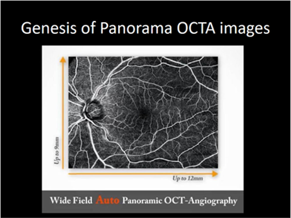
Figure 2:
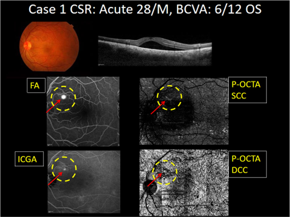
Figure 3:
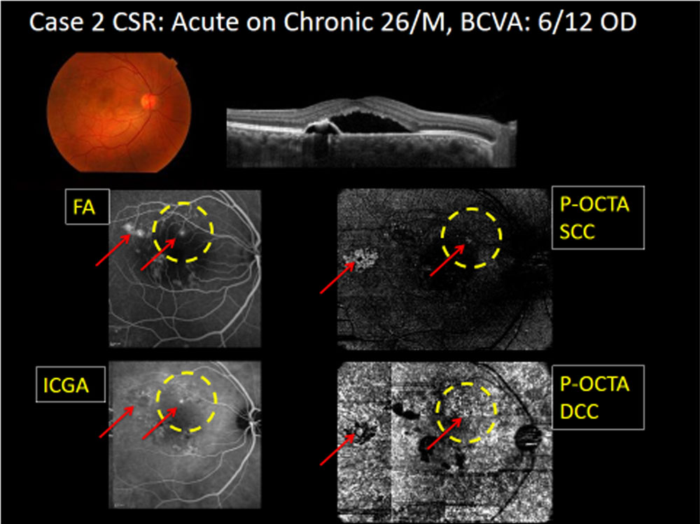
Figure 4:
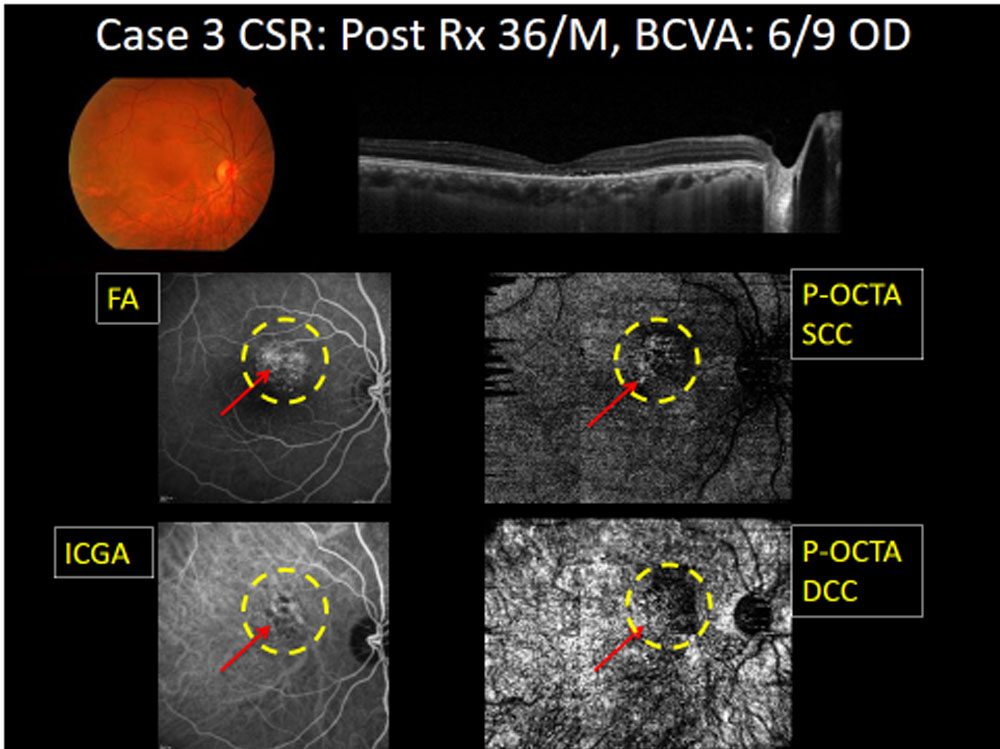
Figure 5:
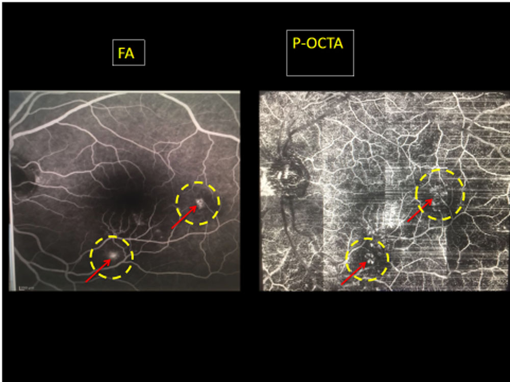
Figure 6:
Leave a Comment