

Dr.Pradeep Kumar, Dr.Vijay Mathur, Dr.Bhupesh Bhatkoti, Dr.Ishan Aggarwal
ABSTRACT
Aim:
Systemic steroid therapy increases choroidal thickness and induces pachychoroid. Prospective, descriptive study.Materials and Methods: Consecutive patients with non-ophthalmological diseases being placed on systemic steroid therapy were enrolled. Choroidal thickness was measured on Day 0 and subsequently at 2 week, 6 week, 12 week and 24 week follow up.
Outcome parameters: Net change in choroidal thickness and appearance of pachychoroid vessels. Results: N= 32 patients completed 24 week follow up. A statistically significant correlation was observed between increase in choroidal thickness and systemic steroid therapy at 6, 12 and 24 week follow up. Appearance of pachychoroid vessels in 14/32 eyes and choriocapillaris compression in 8/32 eyes. Conclusion: Systemic steroid therapy increases choroidal thickness and induces appearance of pachychoroid vessels and choriocapillaris compression. This evidence supports the hypothesis of choroidal vascular dysregulation due to steroids.
INTRODUCTION
Pachychoroid spectrum of disease was described in the present decade (1) and saw an explosion of articles describing choroidal changes of pachychoroid (2). This was largely due to improved choroidal imaging (3). Observation of choroidal thickness and morphology gained validation with increasing number of observations made on associations with as yet unrelated entities (4).
The pachychoroid spectrum includes central serous chorioretinopathy (CSCR)(5). Multiple risk factors have been described for occurrence of CSCR. The pathogenesis has been strongly linked with the effect of adrenocorticoid and mineralocorticoid group of hormones (6)(7). The pathway of affection has been described to be the vascular changes induced by these hormones. The final common pathway has been observed as the dilation of choroidal vasculature and increase in choroidal thickness, ie. the pachychoroid(8).
We realized that evidence existed between CSCR, choroidal thickness and increase of ACTH(9), corticosteroids(10), testosterone(11) and hormones used for oral contraception(12). Again there is definiote evidence between pachychoroid and CSCR. We realized a knowledge gap existed in studying the impact of steroids on an undiseased choroid and production of pachychoroid by steroid exposure.
MATERIALS AND METHODS:
Design: Prospective, Descriptive
Consecutive patients visiting the hospital for non-ophthalmological diseases and being placed on systemic steroids for long term (more than 30 days prescription) were identified from the pharmacy registry. These patients were contacted telephonically and requested for an interview in the ophthalmology OPD. During ophthalmologic consultation they were explained about the proposed study and volunteers agreeing to optical coherence tomography examinations on scheduled basis till the end of therapy or 24 weeks, whichever was later, were enrolled. Choroidal thickness was measured on Day 0 and subsequently at 2 week, 6 week, 12 week and 24 week follow up. The study adheres to the tenets of the declarations of Helsinki and to institutional guidelines for research. Informed consent was obtained from all the patients.
Inclusion criteria:
- Prescribed systemic steroids for more than 30 days
- Willing to undergo monthly OCT examination
- Presence of clear media
- Minimum follow up of 24 weeks
Exclusion criteria:
- Presence of any ophthalmologic disease
- Media opacity obstructing OCT capture
- Presence of any systemic disease associated with altered choroidal thickness
All patients underwent careful clinical evaluation including general, systemic and ophthalmic evaluation. Patients were subjected to optical coherence tomography (Cirrus HD, Carl Zeiss Meditec, Jena, Germany). Central subfoveal choroidal thickness (CSCT) was measured after switching on the enhanced depth-imaging mode. The images captured were manually segmented to obtain the choroidal thickness. Three independent observers took the measurements and the mean was calculated for each patient. Further the vessels were analyzed for dilated choroidal vessel appearance by all three observers and pachy vessels were considered present if two or more observers agreed.
The datawas tabulated using Microsoft excel and underwent statistical analysis using EpiInfo statistical software. Patients completing min 24 weeks observationwere analyzed.
RESULTS:
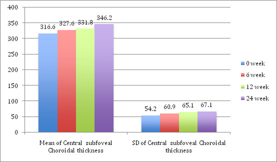
32 patients completed 24 week follow up. The mean CSCT at baseline was 316.6 +/- 54.2 microns. A statistically significant correlation was observed between increase in choroidal thickness and systemic steroid therapy at 6 (327.6 +/- 60.9), 12 (331.8 +/- 65.1) and 24 week (346.2 +/- 67.1) follow up.
Choroidal vasculature was studied for dilated vessels in sattler’s and haller’s layer and appearance of pachychoroid vessels in 14/32 eyes (43.75 %) and choriocapillaris compression in 8/32 (25%) eyes.
DISCUSSION
The changes in choroidal vasculature and thickness have been documented to occur with alteration of steroids (13)(14). This has been studied chiefly in diseases with endogenous hypercortisolism. We studied the effect of steroids on choroid in a prospective cohort.
he mean choroidal thickness has been estimated at 307 +/- 79 microns (15). The subject patients were exposed to systemic steroids for more than 24 weeks at initial doses of nearly 1mg / kg for a minimum prescription of 30 days followed by continuation / gradual tapering up to 24 weeks. This continuous exposure to cortisols was significantly correlated with alterations in choroid in the form of increased SFCT and appearance of pachychoroid vessels. The increased SFCT with exogenous continuous steroids is being reported for the first time.
The development of pachychoroid however does not depend on cortisol exposure alone. There is literature evidence of pachychoroid association with sex (11), daily morning light exposure (16), serum insulin growth factor (17), and sildenafil (18). Thus the study leaves many unanswered questions, while confirming what retinologists have believed for a long time, ie. Steroid exposure can produce pachychoroid. As a corollary, it provides support to the hypothesis of mineralocorticoid antagonism to treat CSCR (19).
LIMITATIONS
The study did not consider the development of CSCR or other ophthalmological impacts of systemic steroids while collecting data.
CONCLUSION
The study provides support to the long believed hypothesis of pachychoroid production with continuous steroid exposure. This strengthens support for systemic mineralocorticoid antagonists in treating CSCR.
REFERENCES
- Gallego-Pinazo R, Dolz-Marco R, Gómez-Ulla F, Mrejen S, Freund KB. Pachychoroid diseases of the macula. Med Hypothesis DiscovInnovOphthalmol J. 2014;3(4):111–5.
- Cheung CMG, Lee WK, Koizumi H, Dansingani K, Lai TYY, Freund KB. Pachychoroid disease. Eye Lond Engl. 2018 Jul 11;
- Ayachit G, Ayachit A, Nadgir H, Joshi S. Validating the pachychoroid disease spectrum using multimodal imaging. Indian J Ophthalmol. 2018;66(7):1022–4.
- Ferro Desideri L, Barra F, Ferrero S, Vagge A. Choroidal thickness changes measured by enhanced depth imaging optical coherence tomography in third trimester pregnant women. Int J Retina Vitr. 2018;4:14.
- Manayath GJ, Shah VS, Saravanan VR, Narendran V. POLYPOIDAL CHOROIDAL VASCULOPATHY ASSOCIATED WITH CENTRAL SEROUS CHORIORETINOPATHY: Pachychoroid Spectrum of Diseases. Retina Phila Pa. 2018 Jun;38(6):1195–204.
- Ambiya V, Goud A, Rasheed MA, Gangakhedkar S, Vupparaboina KK, Chhablani J. Retinal and choroidal changes in steroid-associated central serous chorioretinopathy. Int J Retina Vitr. 2018;4:11.
- Yang D, Eliott D. Systemic Mineralocorticoid Antagonists in the Treatment of Central Serous Chorioretinopathy. SeminOphthalmol. 2017;32(1):36–42.
- Cheung CMG, Lai TYY, Ruamviboonsuk P, Chen S-J, Chen Y, Freund KB, et al. Polypoidal Choroidal Vasculopathy: Definition, Pathogenesis, Diagnosis, and Management. Ophthalmology. 2018 May;125(5):708–24.
- Karaca C, Karaca Z, Kahraman N, Sirakaya E, Oner A, Mirza GE. IS THERE A ROLE OF ACTH IN INCREASED CHOROIDAL THICKNESS IN CUSHING SYNDROME? Retina Phila Pa. 2017 Mar;37(3):536–43.
- Honda S, Miki A, Kusuhara S, Imai H, Nakamura M. CHOROIDAL THICKNESS OF CENTRAL SEROUS CHORIORETINOPATHY SECONDARY TO CORTICOSTEROID USE. Retina Phila Pa. 2017 Aug;37(8):1562–7.
- Cheong KX, Tan CS. Sex-dependent choroidal thickness differences in healthy adults: a study based on original and synthesized data. Curr Eye Res. 2018 Oct 3;
- Madendag Y, Acmaz G, Atas M, Sahin E, Tayyar AT, Madendag IÇ, et al. The Effect of Oral Contraceptive Pills on the Macula, the Retinal Nerve Fiber Layer, and Choroidal Thickness. Med SciMonitInt Med J ExpClin Res. 2017 Nov 27;23:5657–61.
- Wang E, Chen S, Yang H, Yang J, Li Y, Chen Y. CHOROIDAL THICKENING AND PACHYCHOROID IN CUSHING SYNDROME: Correlation With Endogenous Cortisol Level. Retina Phila Pa. 2017 Dec 20;
- Karahan E, Zengin MO, Aydin R, Ozturk T, Kaya M, Kocak N, et al. Correlation of choroidal thickness with serum cortisol level. ClinExpOptom. 2015 Jul;98(4):362–5.
- Akhtar Z, Rishi P, Srikanth R, Rishi E, Bhende M, Raman R. Choroidal thickness in normal Indian subjects using Swept source optical coherence tomography. PloS One. 2018;13(5):e0197457.
- Read SA, Pieterse EC, Alonso-Caneiro D, Bormann R, Hong S, Lo C-H, et al. Daily morning light therapy is associated with an increase in choroidal thickness in healthy young adults. Sci Rep. 2018 May 29;8(1):8200.
- Zhang X, Ma J, Wang Y, Li L, Gao L, Guo X, et al. Elevated serum IGF-1 level enhances retinal and choroidal thickness in untreated acromegaly patients. Endocrine. 2018;59(3):634–42.
- Berrones D, Salcedo-Villanueva G, Morales-Cantón V, Velez-Montoya R. Changes in Retinal and Choroidal Vascular Blood Flow after Oral Sildenafil: An Optical Coherence Tomography Angiography Study. J Ophthalmol. 2017;2017:7174540.
- Willcox A, Culliford L, Ellis L, Rogers CA, Cree A, Chakravarthy U, et al. Clinical efficacy of eplerenone versus placebo for central serous chorioretinopathy: study protocol for the VICI randomised controlled trial. Eye Lond Engl. 2018 Sep 7;
TABLES
Table No 1: Mean and SD changes in choroidal thickness after steroid exposure
Leave a Comment