

Dr.Bharathi bavaharan, Dr.Poornachandra B,Dr.Devanshi Bhanushali,Dr.Rohit Shetty
Abstract:
Aim :
To study the characteristics of photoreceptors in photic retinopathy on rtx1 adaptive optics(AO) and its correlation to visual acuity and the defect seen on conventional spectral domain OCT.
Introduction:
Photic/solar retinopathy is a rare form of retinal injury that can occur due to sun gazing during eclipse or in normal daylight hours. Patients can present with central scotoma sometimes large enough to cause permanent visual loss or remain asymptomatic.Clinically seen as a yellowish lesion, foveal depression or lamellar hole, OCT clinches the diagnosis by a characteristic hyporeflective space in outer retina .The features of photic retinopathy on SD optical coherence tomography (OCT), fundus autofluorescence, microperimetry and fluorescein angiography have been described in previous reports.In vitro studies suggest that the foveal photoreceptor outer segments and retinal pigment epithelium is the primary site of pathology.In vivo visualization of such fine details is now possible with Adaptive optics, that allows clinicians to examine microscopic structures of the eye in living patients with extreme detail in real time.
Till date what we already have is SLO based imaging like time domain OCT, ERG, fundus autofloresence such that we are able to see each layer of retina individually but the next step ahead is seeing each cell individually . AO has utilized technology similar to the way an astronomical telescope is used to view planets by means of reducing the atmospheric aberrations. It aims at correcting the deformations of an incoming wavefront by deforming a mirror in order to compensate for the distortion. So by removing the aberrations in front portion of eye i.e. the tears ,cornea, lens we were able to see the retinal cells and study the changes in photic retinopathy.
Materials and methods:
6 eyes of 5 patients with photic retinopathy were included.Patients with clear media and characteristic defect on SD OCT and low vision not attributable to any other cause were included. Subjects with any other ocular pathology were excluded from the study.All patients underwent thorough clinical examination including best corrected visual acuity (BCVA), cycloplegic refraction slit lamp examination, fundus examination, SD-OCT (Spectralis OCT; Heidelberg Engineering, Heidelberg, Germany ) and adaptive optics (rtx1 adaptive optics imaging) . Informed consent was obtained from the patients after a full explanation of the procedures. All studies were conducted in accordance with the principles embodied in the Declaration of Helsinki.
Results:
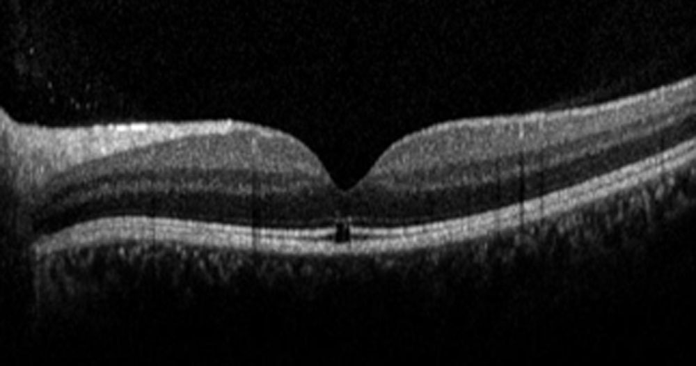
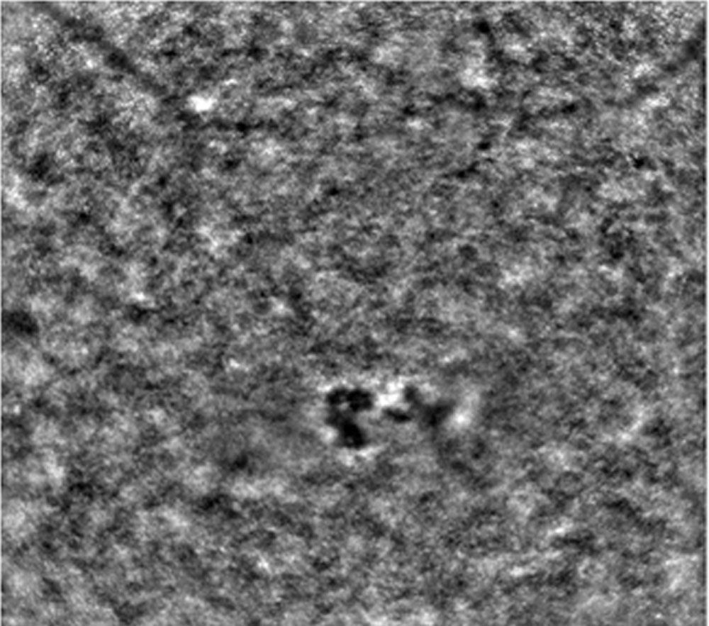
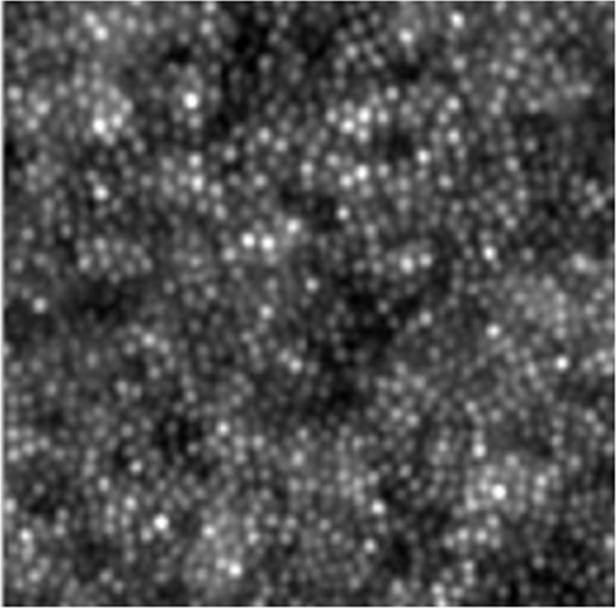
The Mean Age of the patients was 49 ± 18 ( range:23-65).There were 3 Males and 2 Females . Area affected was slightly larger than the defect seen on routine standard SD OCT(Figure 1).Cone count was found to significantly decrease at 0.75 and 1 degree (p < 0.05). However it was preserved beyond 1 degree(Table 2,fig 2).Increase in spacing was noticed at 0.75 and 1 degree as compared to normal.Cone spacing was noted close to normal beyond 1 degree (Table 3,figure 2) Cone mosaic was preserved beyond 1 degree in all patients suggesting the focal nature of the defect.Larger defect corresponded to poor vision in all cases.Sub group analysis showed patients with good cone count had better prognosis and chances of earlier recovery.Adaptive optics shows decreased cone count larger than the defect seen on SD-OCT suggesting a possible diffuse phenomenon which is probably answer to the varying vision loss and recovery in patients with similar defect .
Discussion
Photic/Solar retinopathy is results from direct sungazing and is usually reported following eclipse viewing. Proposed mechanisms of injury include photochemical toxicity generated by excess short-wavelength visible light and thermal injury from near-infrared radiation.In the acute stage,a yellow-white spot is seen in the fovea, which corresponds to a hyperreflective photoreceptor bands on OCT.Later the yellow-white spot may fade or evolve into a reddish cyst like lesion with surrounding mottled pigmentation that appears as hyporeflectivity of photoreceptor bands on OCT.During a partial solar eclipse, when part of the sun’s core remains visible, viewing the solar rim without eclipse viewing glasses which have special-purpose solar filters can lead to severe solar retinopathy
with an absolute central scotoma.Adaptive optics provides cellular level images of the disturbed photoreceptor mosaic.In our study we found that the cone mosaic was preserved beyond 1 degree in all patients suggesting the focal nature of the defect.Larger defect corresponded to poor vision in all cases.Sub group analysis showed patients with good cone count had better prognosis and chances of earlier recovery. Adaptive optics shows decreased cone count larger than the defect seen on SD-OCT . Limitations were the smaller sample size and lack of longitudinal follow up.
Conclusion
Adaptive optics imaging enhances the resolution of retinal imaging devices by correcting ocular-induced wavefront aberrations, allowing in vivo visualization of cellular structures .It shows the defect in photoreceptors at larger area than conventional SD OCT. It is a senstive and accurate tool to measure the cones in solar maculopathy.It can be used to predict and observe changes in these patients over time.Helps us in better understanding of pathogenesis and prediction of prognosis .
Table 1
| BCVA | Defect in OCT in micrometre | |
| Case 1 | 6/9 | 143 |
| Case 2 | 6/18 | 147 |
| Case 3 | 6/24 | 175 |
| Case 4 | 6/9 | 83 |
| Case 5 | 6/12 | 187 |
| Case 6 | 6/24 | 190 |
Table 2
| Cone count | Solar retinopathy | Normal |
| 0.75 degree | 14352 | 25693 |
| 1 degree | 15891 | 26873 |
| 2 degree | 27601 | 27726 |
Table 3
| Cone spacing | Solar retinopathy | Normal |
| 0.75 degree | 14.13 | 5.34 |
| 1 degree | 12.57 | 5.67 |
| 2 degree | 6.27 | 6.16 |
Leave a Comment