

Dr.Rajeshgoyal, Dr. JUGAL KISHORE CHOUHAN,Prof.Pankaj Sharma
Introduction
In 2006, Melles introduced Descemet’s Membrane Endothelial Keratoplasty (DMEK) which involved descemet’s membrane and endothelial layer transplantation after removal of host descemet’s membrane. 1
Descemet Membrane Endothelial Keratoplasty (DMEK) offers better & faster visual rehabilitation and a lower risk of immune rejection than Penetrating Keratoplasty (PK) and Descement membrane Endothelial Keratoplasty(DSEK).2-6
DMEK is replacing DSEK and is becoming the preferred procedure for treatment of endothelial dysfunction . 7,8DMEK is more technically difficult than DSEK and it has a steeper learning curve . 9 -11.
DMEK offers a two step challenge to the surgeon. One is to do meticulous stripping of donor’s descemet’s membrane and then performing intraocular donor graft manipulation. It is important to strip the donor’s descemet’smembrane completely without leaving any remnant behind. This is important as it interferes with optical performance and adhesion of the graft. It is important to stain the graft properly and avoid over staining. Marking of the graft on descemet’s side should be done .Graft tissue should be made in double roll and before injecting, the orientation of the graft should be checked. Folding of DMEK graft occurs in a fashion that the endothelial side always remains out. Unfolding of the donor DMEK tissue in the host eye is the single most important step for successful DMEK. For successful graft attachment, posterior surface of the recipient cornea should be cleared of descemet’s membrane but over enthusiastic roughening should be avoided. Air bubble is used to attach the graft. Too much big air bubble is avoided as it may increase the pressure in the eye and in turn damage the cell population.
Due to shortage of corneal donors, the possibility of using one donor cornea for preparing descemet’s membrane graft for DMEK and remaining tissue for Deep Anterior Lamellar Keratoplasty (DALK) is being explored. There is also temptation to split a single descemet’s membrane for use in two or even four patients.
Aims & Objectives
Prospectively evaluating the 6 months results of Descemet Membrane Endothelial Keratoplasty done for management of endothelial decompensation .
Material & Methods
The study was conducted at Upgraded Department of Ophthalmology , SMS MEDICAL COLLEGE and Hospital ,Jaipur and all the cases were operated by a single surgeon .
It was a prospective study done between June 2017 to Dec 2017 with a follow up period of 6 months .
Inclusion Criteria (12-17)
- Fuch’s endothelial dystrophy
- Bullous keratopathy (both pseudophakic and aphakic)
- Posterior polymorphous corneal dystrophy
- Congenital hereditary endothelial dystrophy
- Iridiocorneal endothelial (ICE) syndrome
- Previous failed PK or EK
- Post traumatic endothelial dystrophy
- Post recurrent herpetic endothelitis.
Exclusion Criteria
1.Anterior stromal scarring
2.Full thickness corneal opacity
3.Adherent leucoma
4.High irregular astigmatism.
DMEK was performed in 40 eyes with endothelial decompensation . Best spectacle corrected visual acuity (BCVA) and endothelial cell density (ECD) were evaluated before and at 1, 3, and 6 months after surgery. Intra- and postoperative complications were documented.
Surgical Steps
Graft Preparation
Corneal graft was mounted on the Teflon block with endothelial side down and graft was marked in the centre of the cornea . Then graft was inverted so that endothelium was now facing upward . Now Trypan Blue dye 0.06% was injected over the graft so as to visualize the endothelium .
After that using 10 mm simple corneal trephine a gentle mark was made on the cornea. Using sinskey endothelium Descemet’s complex was separated from peripheral 360degree . Again now graft was punched with 8mm corneal trephine so as to make a partial thickness cut . Using a plain forcep Descemet’s Endothelium complex was separated from stroma of the Donor cornea leaving small attachment at the periphery.
On the bare stroma 3mm punch was made ; endothelium was again reposited on the stroma . Graft was reinverted and a F mark was made on the Descemet’s side so as to recognize correct orientation of graft inside the eye .
Graft was separated from stroma and trypan blue dye 0.06% was placed over the graft for 1 minute so as to stain the graft subsequently, DM was
placed in balanced salt solution in a glass dish while the DM of the recipient
was stripped.
Graft was aspirated with a glass cartridge attached to a 5 cc syringe .
Surgery
All surgeries were performed under peribulbar block .
After painting & draping the patient under aseptic condition , 2 paracentesis were made at 6 & 12 o’ clock position . If the epithelium was edematous it was scrapped off using 15 no blade .
2.8 mm temporal incision was made using a keratome . If the cataract surgery was combined with DMEK first phaco with IOL was done and then Endothelium was stripped from the host using reverse sinskey .
Peripheral iridotomy was made at 6 o’ clock position using a vitrector . A/C maintainer was placed at 6 o’ clock position . As soon as glass cartridge was placed inside the eye ; A/C maintainer was stopped and graft was injected inside the eye with graft in a double roll configuration .
Keeping anterior chamber shallow and using iris as 3rd hand graft was properly oriented with endothelium facing down .
Now air was injected in the anterior chamber so as to appose the graft to the stroma . BCL was placed over the eye . After instilling antibiotics drops eye was patched . The patient was instructed to stay in the supine position overnight .
Results
Average age of the recipient was 68 yrs (Ranging from 50 to 86 yr age).
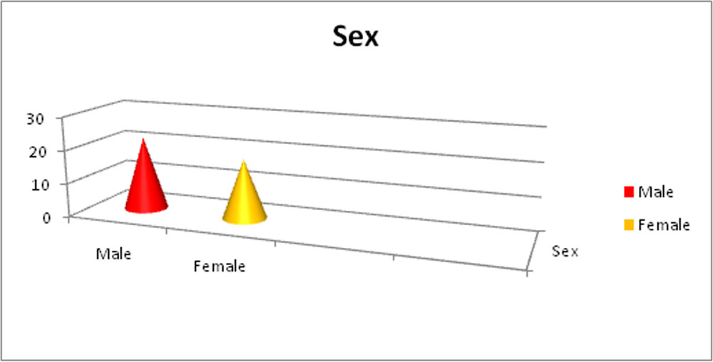
There were 22 males & 18 females .
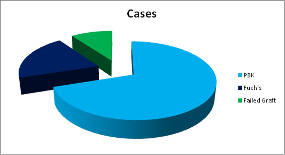
Indications of DMEK were Pseudophakic Bullous Keratopathy 28 cases , Fuch’s Dystrophy 8 cases and Failed Keratoplasty 4 cases .
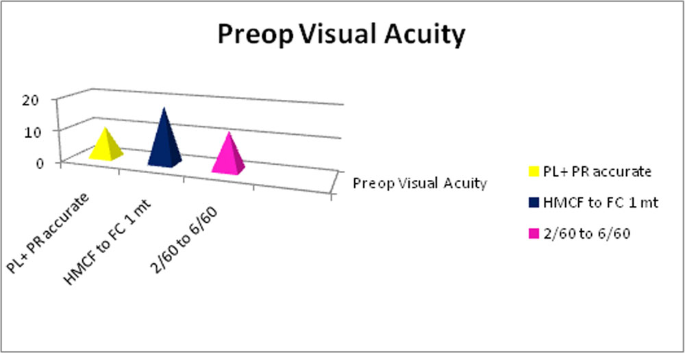
Preoperative visual acuity ranged from Pl + PR Accurate in 10 cases . Hand movement close to face to finger counting 1 meter 18 cases & 2/60 to 6/60 in 12 cases
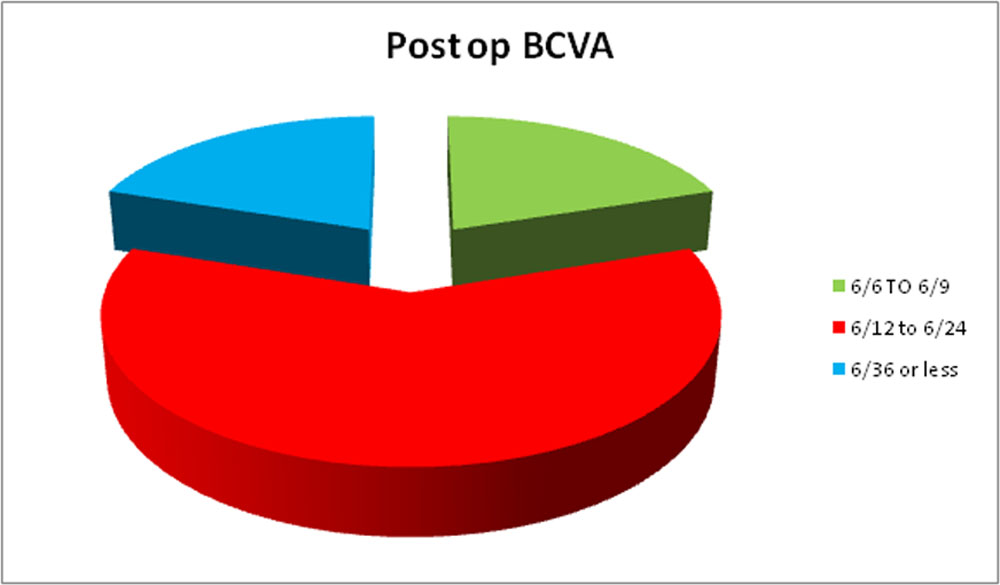
At 6 months Postoperative Best Corrected Visual Acuity ranged from 6/6 to 6/9 (8 cases), 6/12 to 6/24 (24 cases ) & 6/36 or less (8 cases)
Potential vision limiting factors included corneal scar 4 cases , Glaucoma 2 cases , ARMD 1 case & Cystoid Macular Edema 1 case .
35 cases were performed as a single procedure & 5 cases were performed as DMEK with Cataract extraction & PCIOL implantation .
38 patients had clear graft and 2 patients had Primary graft failure and they underwent DSEK.
Average Donor Endothelial Cell Density showed a decrease from 2610(± 100) cells/mm2 before to 1504 (±100)cells/mm2at 6 months after surgery.
Refractive stability was shown at 3 months after DMEK, i.e. no significant change in refractive cylinder at 3 versus 6 months of follow-up. The main complication was (partial) graft detachment occurring in 15% eyes . Secondary ocular hypertension was seen in 10% eyes. Secondary cataract requiring phaco-emulsification developed in 2 eyes.
Discussion
DMEK is technically demanding because the delicate thin donor tissues tend to fold into a tight scroll. Many techniques have been described to facilitate the unfolding of the DM scroll. Use of corneas from older donors is preferred. It has been reported that donors aged >65 years had a greater graft stiffness, which facilitates the unfolding process. A few techniques have been developed to successfully unfold the DM grafts and reduce the need for air injection. These techniques include elimination of viscoelastic, use of SF6, and oversizing
the descemetorrhexis of the recipients. Often, a combination of surgical techniques are required to efficiently unfold the DM scroll .
Partial detachment was the most common complication in the present study. This complication was managed successfully with a single air injection in the anterior chamber to facilitate visual recovery .
There are several limitations to the current study. First ,this study was a small prospective evaluation of only 40 patients. Second, the follow-up period was relatively short. Therefore the results reflect the short term outcome. Third, the current series was from a single center. A larger series of patients treated by different surgeons at multiple centers would be necessary to further evaluate the overall outcome of DMEK.
CONCLUSION
DMEK may provide a refractively neutral near complete, rapid visual rehabilitation with ECDs similar to earlier endothelial keratoplasty techniques. This combined with a relatively low complication rate, would indicate that DMEK is a safe and effective treatment for corneal endothelial disorders. Even with so many advantages, DMEK still remains in limited surgical hands due to its steep learning curve.(18-19)
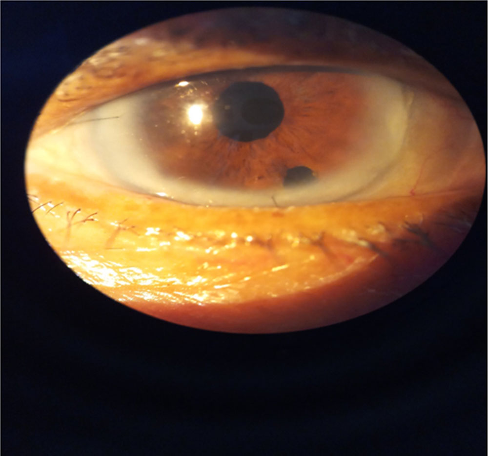
1 WEEK POST OPERATIVE PICTURE OF DMEK SURGERY – SMS HOSPITAL
References :
- Droutsas K, Giallouros E, Melles G, Chatzistefanou K, Sekundo W.Descemet’s membrane endothelial keratoplasty: Learning curve of a single surgeon. Cornea 2013;32:1075-1079
- Anshu A, PriceMO, Price FW Jr. Risk of corneal transplant rejection significantly reduced with Descemet’s membrane endothelial keratoplasty. Ophthalmology 2012;119(3):536–540.
- Van Dijk K, Ham L, Tse WH, et al. Near complete visual recovery and refractive stability in modern corneal transplantation: Descemet membrane endothelial keratoplasty (DMEK).Cont Lens Anterior Eye 2013;36(1):13–21.
- Chaurasia S, Price FW Jr, Gunderson L, Price MO. Descemet’s membrane endothelial keratoplasty: clinical results of single versus triple procedures combined with cataract surgery).Ophthalmology 2014;121(2):454–458.
- Feng MT, Price MO, Miller JM, Price FW Jr. Air reinjection and endothelial cell density in Descemet membrane endothelial keratoplasty: five-year follow-up. J Cataract Refract Surg2014;40(7):1116–1121.
- Dapena I, Ham L, Netukova M, van der Wees J, Melles GR.Incidence of early allograft rejection after Descemet membrane endothelial keratoplasty. Cornea 2011;30(12): 1341–1345
- Tourtas T, Laaser K, Bachmann BO, Cursiefen C, Kruse FE.Descemet membrane endothelial keratoplasty versus descemet stripping automated endothelial keratoplasty. Am J Ophthalmol 2012;153(6):1082–1090.e1082.
- 8.Kruse FE, Schrehardt US, Tourtas T. Optimizing outcomes with Descemet’s membrane endothelial keratoplasty. Curr Opin Ophthalmol 2014;25(4):325–334.
- Tenkman LR, Price FW, Price MO. Descemet membrane endothelial keratoplasty donor preparation: navigating challenges and improving efficiency. Cornea 2014;33(3):319–325.
- Dapena I, Ham L, Droutsas K, van Dijk K, Moutsouris K,Melles GR. Learning curve in Descemet’s membrane endothelial keratoplasty: first series of 135 consecutive cases. Ophthalmology 2011;118(11):2147–2154.
- Monnereau C, Quilendrino R, Dapena I, et al. Multicenter Study of Descemet Membrane Endothelial Keratoplasty: First Case Series of 18 Surgeons. JAMA Ophthalmol 2014;132(10): 1192–1198.
- Price FW Jr., Price MO. Descemet’s stripping with endothelialkeratoplasty in 50 eyes: A refractive neutral corneal transplant.J Refract Surg 2005;21:339‑45.
- Eye Bank Association of America. Eye Banking Statistical Reports.Washington, DC: Eye Bank Association of America; 2005‑2011.
- Anshu A, Price MO, Tan DT, Price FW Jr. Endothelial keratoplasty:A revolution in evolution.SurvOphthalmol 2012;57:236‑52.
- Duman F, Kosker M, Suri K, Reddy JC, Ma JF, Hammersmith KM,et al. Indications and outcomes of corneal transplantation ingeriatric patients. Am J Ophthalmol. 2013;156:600-7.e2.
- Tan JC, Holland SP, Dubord PJ, Moloney G, McCarthy M,Yeung SN. Evolving indications for and trends in keratoplasty inBritish Columbia, Canada, from 2002 to 2011: A 10‑year review.Cornea 2014;33:252‑6.
- Guerra FP, Anshu A, Price MO, Giebel AW, Price FW. Descemet’smembrane endothelial keratoplasty: Prospective study of1‑year visual outcomes, graft survival, and endothelial cell loss.Ophthalmology 2011;118:2368‑2373.
- Hamzaoglu EC, Straiko MD, Mayko ZM, Sales CS, Terry MA.The first 100 eyes of standardized Descemet strippingautomated endothelial keratoplasty versus standardized
- 19.Green M, Wilkins MR, Comparison of early surgical experience andVisual outcomes of DSAEK and DMEK. Cornea 2015;34:1341‑1344
Leave a Comment