

Dr.Visakh Thomas, Dr.Niveditha Narayanan
ABSTRACT
Purpose:
To describe the outcomes of LS-LK for the management of advanced corneal ectasia.
Methods:
A retrospective case series from 3 eyes of 3 patients with acquired keratoglobus, two of them being one eyed and one with Pellucid Marginal Degeneration progressing into keratoglobus and 3 eyes of 2 patients with congenital keratoglobus and Ehlers-Danlos syndrome, out of which one presented with trauma and globe rupture. Visual outcomes and anatomical results(corneal thickness and keratometry)were evaluated.
Results:
Mean follow up was 1year.In all patients corneal thickness increased and central keratometry decreased. All patients except one benefited, with visual acuity improving by more than 2 lines within a period of 6 months.
Conclusions:
Prompt diagnosis and early intervention with LSLK is a viable option for improving the visual acuity and preserving ocular integrity in patients with advanced corneal ectasia, before the occurrence of vision-threatening traumatic corneal rupture.
Keywords
Keratoglobus, limbal stem cell sparing, lamellar keratoplasty
INTRODUCTION
Keratoglobus (KGB) is a rare, noninflammatory corneal thinning disorder characterised by generalised thinning and globular protrusion of the cornea1. It can be congenital and acquired. The congenital form is always bilateral and is associated with Ehlers–Danlos syndrome type VI, Lebers congenitalamaurosis,, the blue sclera syndrome, Marfan syndrome, and Rubinstein–Taybi syndrome and osteogenesis imperfecta.2 The acquired form, which presents in adulthood, may evolve from preexisting pellucid marginal degeneration (PMD) or keratoconus.3
Surgery is indicated when visual needs are no longer satisfied, or when there is a risk of occurrence of corneal rupture.4 The surgical aim should be to restore corneal and scleral anatomical integrity by replacing or supporting weak corneal tissue, both peripherally and centrally, while maintaining central corneal optical clarity.5 Here, we describe a surgical technique of limbal sparing lamellar keratoplasty(LS-LK) for the management of advanced corneal ectasia like keratoglobus.
METHODS
In a retrospective case series, six eyes of five patients including 3 female and 2 male subjects whose age ranged from 11 to 32 years were operated. Two patients were one eyed, with a history of trivial injury being the cause of their blind eye. Three patients had acquired keratoglobus, among them, two had pallucid marginal degeneration progressing into keratoglobus and two patients with congenital keratoglobus along with Ehler Danlos syndrome, out of which one presented with trauma and corneal rupture .
Before operation, all the patients had documented progressive corneal thinning(evaluated by OCULUS Pentacam ® ).The study was conducted from October 2011 to June 2018.
Surgical technique
All the procedures were performed under general anesthesia. Using an appropriate sized corneal trephine the host cornea was marked 1.5-2 mm inside the limbus.Using a crescent knife, a 360 degree corneal stromal pocket was made in the periphery,preserving the limbal stem cells. Corneal epithelial debridement was performed with ethanol,which was thoroughly rinsed with plenty of Balanced salt solution. Radial conjunctival incision was made in each of the 4 quadrants and tenons dissected 360-degree.Corneal periphery along with limbus and conjunctiva cut and retracted to form four flaps.A free hand scleral ridge was made around the
cornea. The donor tissue was prepared from a fresh whole globe or a corneal button.The margins of the donor tissue were trimmed cirumferentially to create a tapered edge for better stability and apposition. Donor descemets membrane was stained with trypan blue and then peeled off. Donor size ranged from 12 to 14 mm.The donor graft was positioned and fixed to host sclera using 16-24 interrupted 10-0 nylon sutures.Thereafter, the retracted host limboconjunctival tissue was repositioned and fixed with glue 360 degrees. Eye is then patched with steroid drops.
Postoperative Management
The patients were closely monitored every alternate day till complete epithelisation was noted. Then patients were examined weekly for 1 month , biweekly for 3 months, monthly till 6 months and then 3 monthly till the last follow-up. Among them the longest follow-up was of 4 years. Postoperatively,bandage contact lens was applied for all patients along with topical antibiotic agent(Moxifloxacin 0.5%) 6 times a day till the bandage contact lens was removed and full epithelisation was noted. Also on postoperative day1 all patients were prescribed corticosteroid drops (Betamethasone 0.1%) 12 times a day in tapering dose and topical artificial tears(carboxymethyl cellulose sodium) 6 times a day.Loose sutures were removed as and when required.
RESULTS
All patients were followed up for atleast 1 year. There was significant improvement in visual acuity in all the patients(6/9 to 6/18) except one and corneal thickness improved in all patients.
Postoperatively there was no significant interface foreign body or debris but there were two cases each of interface haze and interface haeme .Complete resolution of the interface haeme was achieved but the interface haze persisted. One patient had failed graft and one had to undergo repeated PK(Penetrating Keratoplasty) with Ahmed glaucoma valve implantation due to uncontrolled intraocular pressure.
Case 1
29 year old female presented with recently diagnosed diminision of vision in the right eye for which she was not using glasses. She underwent a comprehensive eye examination which showed her uncorrected vision to be 2/60 in the right eye and 6/36 in the left eye, and best corrected visual acuity to be 6/24 in right eye with -10.50 DS/ -3.50 @ 90 correction and 6/18 in left eye with plano with -4.0 DC @90 correction with the rest of the ocular examination within normal limits.A full ectasia workup was done ,which includedpentacam, topography, ASOCT(Anterior segment Optical Coherence tomography), pachymetry and specular microscopy, which revealed advanced pellucid marginal degeneration with global corneal thinning in the right eye with corneal thickness of 217 μm at the thinnest location. Patient underwent LS-LK for the right eye and in the postoperative period ,patient achieved an increase in corneal thickness and reduction in central keratometry reading from 56.50 D to 47.50 D, which resulted in improved visual acuity to 6/9 with an acceptable correction of -4.0 DS/-4.0 DC @90 . Patient is on regular follow up for 17 months and is maintaining her vision.
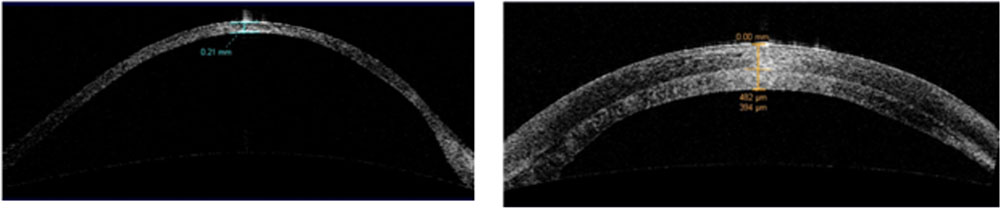
Fig. 1Fig. 2
Fig 1. ASOCT showing preopoperative marked peripheral and central corneal thinning
Fig 2. ASOCT showing good apposition of the host and the graft along with significant increase in corneal thickness
CASE 2
A 24-year-old man presented with severe decreased vision in both eyes. On comprehensive ophthalmic evaluation ,he was found to have an uncorrected visual acuity of counting fingers at 50cm in the right eye and 3/60 in left eye. His vision wasnot improving in the right eye with correction and it improved to 6/36 in the left eye with a correction of -18.0 DS/-3.50 @40. There was apical scaring in both the eyes with global thinning and protrusion of the cornea. Rest of the ocular examination was within normal limits. His ectasia workup showed severe thinning of the cornea with corneal thickness of 147μm in right eye and 218μm in left eye. He was advised LS-LK for the right eye but he was lost to followup. Two years later he was referred here for the left eye LS-LK as he had spontaneous eviseration following a minor trauma in the right eye. Left eye visual acuity has reduced to 3/60 from the last visit and ectasia workup showed progressive thinning and protrusion of the cornea. LS-LK was performed for the left eye and the only post-operative complication was interface haemewhich was self-resolving. Patient is on regular followup for 15 months and he achieved a significant increase in the corneal thickness, reduction in keratometry value from 69.06D to 46.30 D and a significant improvement in the visionto 6/9 with a refractive correction of -2.5DS/ -5.0 DC @ 60.
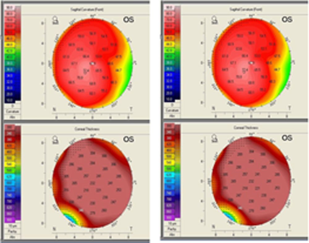
Fig.3 Fig. 4
Fig 3. Corneal topography displaying the preoperative sagital curvature and pachymetry
Fig 4. Corneal topography displaying the postperativesagital curvature and pachymetry, showing the significant increase in corneal thickness and reduction in corneal steepness.
Case 3
A 32-year-old man ,practicallya one eyed patient with only light perception in the left eye,which was due to corneal rupture following a trivial fall. Primary repair and retinal surgery was done elsewhere but there was nill vision improvent. He was a known case ofkeratoglobusin both the eyes and was on RGP (rigid gas permeable)contact lens when this trauma happened. He was referred to our cornea clinic for further management of the right eye. His comprehensive ocular examination showed an uncorrected visual acuity of 6/36 in right eye and light perception in left eye. His best corrected visual acuity was 6/24 with a refractive correction of-1.50 DS /-6.00 DC @100 and no improvement in left eye.On examination he had advanced acquired keratoglobus due to progressive pellucid marginal degeneration which was confirmed by ectasia workup which showed a thinnest pachymetryof 57 μmlocated superiorly. Patient underwent LS-LK in the right eye.Interface haze was noted in the postoperative period which resolved completely with topical steroids.His cornealthickness increased , central 3-mm keratometry decreased from 59.40 to49.62 D and the best corrected visual acuity achieved at the end of 14 months was6/7.5 with a refractive correction of -3.00DS/-6.0 DC @ 60.
Case 4
A 14-year-old girl, elder sister of case 5,born out of consanguineous marriageand diagnosed with Ehlers-Danlos syndrome with blue sclera and joint hyper-extensibility. She underwent a comprehensive eye examination and was found to have a best corrected visual acuity of 6/24 in the right eye with a refractive correction of-9.00DS/-6.00DC @170 and 6/18 in left eye with refractive correction of -6.00 DS/-5.00
DC @ 170.ectasia workup revealed overall thinned cornea in both eyes with thinnest pachymetry of 256μm for the right eye and 290μm forthe left eye. She was advised LS-LK for the right eye which she underwent. Interface haze was noted in the postoperative period which significantly reduced with corticosteroids ,but a faint remnance being noted at the last follow up which was 3 years post operatively. Her vision improved from 6/24 to 6/9 with a correction of ± / -3.50 DC@180, along with a significant increase in corneal thickness and reduction in central 3-mm keratometryreading from 63.39 to 45.30 D.
Case 5
A 11-year-old girl, elder sister of case 4 born out of consanguineous marriage and diagnosed with Ehlers-Danlos syndrome with blue sclera and joint hyper-extensibility presented with severely decreased vision in both eyes.She underwent a comprehensive ophthalmic examination and was found to have an uncorrected visual acuity of 6/60 in both eyes improving to 6/45 in each eye with a refractive correction of-7.00DS/ -5.50 DC @ 160 for the right eye and -9.25DS/-7.00DC @170 for the left eye.Ectasia workup revealed overall thinned cornea in both eyes with thinnest pachymetry values were 247 μm and 250 μm for right and left eye respectively.She was advised LS-LK for both the eyes but during this waiting period for the surgerya trivial injury with finger nail resulted incorneal rupture with lens extrusion .Primary repair was done but with no success, so the patient was immediately taken up for temporary LK-LS for tectonic support along with vitrectomy.Iinterface haze and vascularization was noted postoperatively.At this point her left eye showed progression of the disease and hence was taken up for LS-LK.Meanwhile her right eyevisual acuity improved to 6/24 at glass appointment but there waspersistant interface haze and vascularization and by the end of one year the graft failed.So the patient underwent PK +Vitrectomy +SFIOL(Scleral fixated intra ocular lens).By this time her left eye graft also failed and was taken up for lamellar keratoplasty.Again for the right eye the graft failed along with uncontrolled IOP with maximum glaucoma medication and hence was taken up for re-PK and AGV ( Ahmed glaucoma valve) implantation.Post operatively in both the eyes visual acuity improved to 6/18 with a correction of ± /-4.0 DC @5 in right eye and plano in left eye.
DISCUSSION
Keratoglobus is a non-inflammatoryectatic disorder of the cornea characterized by thinning from limbus to limbus resulting in globular protrusion of the cornea.Cornealtear or globe rupture in keratoglobus can occur either spontaneously or following trivial trauma. Two of our patients with diagnosed keratoglobus were advised surgery but patients were lost to follow-up.During this period one had spontaneous eviseration following a minor trauma and the other had visual acuity of only perception of light following trivial fall indicating the blinding nature of the disease.
Collagen cross linking (CXL) is a good option for stabilizing corneal thickness.Due to its potential of endothelial toxicity and cell death, CXL is contraindicated for individuals with corneas thinner than 350 μm.In our study all the patients had pachymetry reading less than 350 μm, hence it was deferred for them.
Surgical management of keratoglobus is an extremely difficult task.Various techniques have been proposed and traditional keratoplasty was one of the first surgical procedures attempted. Other surgical techniques include LKP6( lamellarkeratoplasty), tuck in LKP 7,8,epikeratoplasty, and corneoscleroplasty. Because of severe generalized corneal thinning and protrusion , PK and LKP are demanding procedures with a high risk of wound leakage, wound instability,malposition,high astigmatism, delayed epithelial healing and recurrence of the ectatic disorder leading to poor visual outcomes. Further , a large diameter PK entails an increased risk of graft rejection, delayed re-epithelisation, and post operative glaucoma due to the proximity of the graft to limbal stem cell, limbal vasculature, as well as the trabecular meshwork. Tuck in LKP is technically demanding because it involves the lamellar dissection of a thin host bed, which may be difficult or sometimes impossible to perform.
In conclusion ,LS-LK restores the structural integrity of the cornea in keratoglobus withoutjeopardizing the limbal stem cells.But it has its own set of drawbacks like interface haze,graft failure ,meticulous long term follow up and a steeplearning curve for the surgeons.
REFERENCES
- Wallang BS, Das S. Keratoglobus. Eye. 2013 Sep;27(9):1004–12.
- Hyams SW, Kar H, Neumann E. Blue sclerae and keratoglobus. Ocular signs of a systemic connective tissue disorder. Br J Ophthalmol. 1969;53(1):53
- Cameron JA, Al-Rajhi AA, Badr IA. Corneal ectasia in vernal keratoconjunctivitis. Ophthalmology. 1989;96(11):1615–1623.
- Hallermann W. Technique and results of an operative treatment of keratoglobus (author’s transl). KlinMonatsblAugenheilkd. 1975;166(5):593–598.
- Jones DH, Kirkness CM. A new surgical technique for keratoglobus-tectonic lamellar keratoplasty followed by secondary penetrating keratoplasty. Cornea. 2001 Nov 1;20(8):885-7.
- Kaushal S, Jhanji V, Sharma N, Tandon R, Titiyal JS, Vajpayee RB. “Tuck In” Lamellar Keratoplasty (TILK) for corneal ectasias involving corneal periphery. Br J Ophthalmol. 2008;92(2):286–290.
- Vajpayee RB, Bhartiya P, Sharma N. Central lamellar keratoplasty with peripheral intralamellar tuck: a new surgical technique for keratoglobus. Cornea. 2002 Oct 1;21(7):657-60.
Leave a Comment